Medical AI · Knowledge-Informed Learning · Clinical Decision Support
Knowledge-Informed Constrained Learning for Data-Efficient Extracapsular Extension Assessment in Head and Neck CT
Under review at Medical Image Analysis
Research problem
Extracapsular extension occurs when cancer spreads beyond the lymph-node capsule. It is an important prognostic factor in head and neck cancer and can influence staging, prognosis, and treatment planning. Preoperative assessment from CT remains difficult because the radiologic signs may be subtle, heterogeneous, and localized around the nodal boundary, while pathology-confirmed labels are limited.
Conventional models may learn from the complete CT volume, including anatomically unrelated structures, or use segmentation only as a preprocessing or auxiliary task. Both strategies leave the classifier vulnerable to irrelevant background features and provide a weak connection between localization and the final clinical prediction.
Knowledge-informed constrained learning
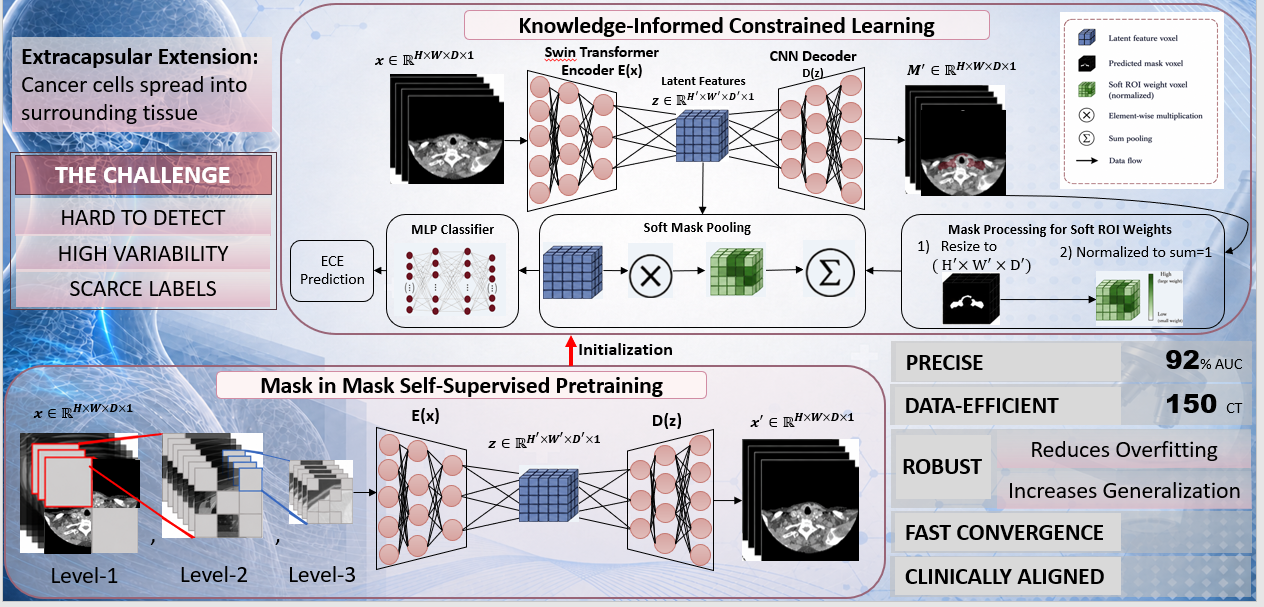
The framework uses a shared dual-output 3D SwinUNETR to jointly predict the lymph-node region and classify ECE. Anatomical supervision therefore shapes the latent representation rather than serving only as a separate preprocessing step.
The predicted anatomical mask is projected into the latent feature space and normalized into soft spatial weights. These weights determine which features reach the classifier, emphasizing nodal and perinodal evidence while suppressing unrelated background anatomy. The model consequently learns both where to look and which evidence should support the final decision.
Clinical prior and data-efficient learning
Because ECE is defined by spread beyond the lymph-node capsule, the supervision target is expanded to include clinically plausible perinodal soft tissue while excluding implausible regions such as air and bone. This incorporates the disease mechanism directly into model training.
To address scarce pathology-confirmed labels, the encoder is initialized through Mask-in-Mask self-supervised pretraining on approximately 1,500 unlabeled head-and-neck CT volumes. The pretraining stage learns anatomical representations at multiple spatial scales before supervised fine-tuning on the ECE cohort.
Selected results
The study evaluated 150 patients with pathology-confirmed ECE status. The proposed model achieved:
- Accuracy: 0.924
- AUC: 0.928
- Sensitivity: 0.926
- Specificity: 0.892
- Lymph-node ROI segmentation Dice: 0.821
The model achieved the strongest accuracy, AUC, sensitivity, and Dice among the evaluated methods. During inference, it predicts the anatomical region directly from the CT volume and uses that prediction to guide classification, so no manually supplied ROI is required for the final decision.
Interpretability and decision support
The soft spatial weights are part of the model's actual decision pathway, not a post hoc explanation. They show where the classifier aggregates evidence, while Grad-CAM provides a complementary analysis of which regions influence the final prediction. Together with the predicted anatomical mask, these outputs make it possible to examine whether the decision is supported by clinically meaningful nodal and perinodal evidence.
Project support
NIH-USA